It's simple
Create a presentation, share it with the community and get hints on possible solutions

Step 1
Create a presentation using our template in Google Slides

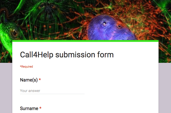
Step 2
Fill the submission form and share your presentation

Step 3
Get the help you need - during a session or later via #call4help on Image.sc forum
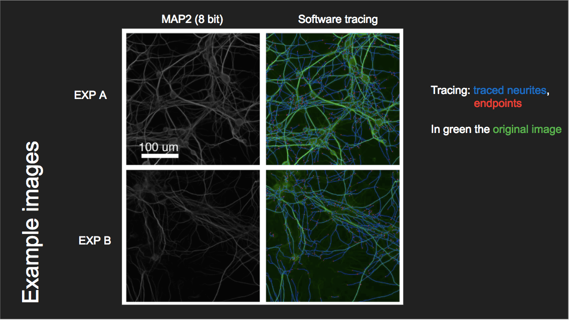
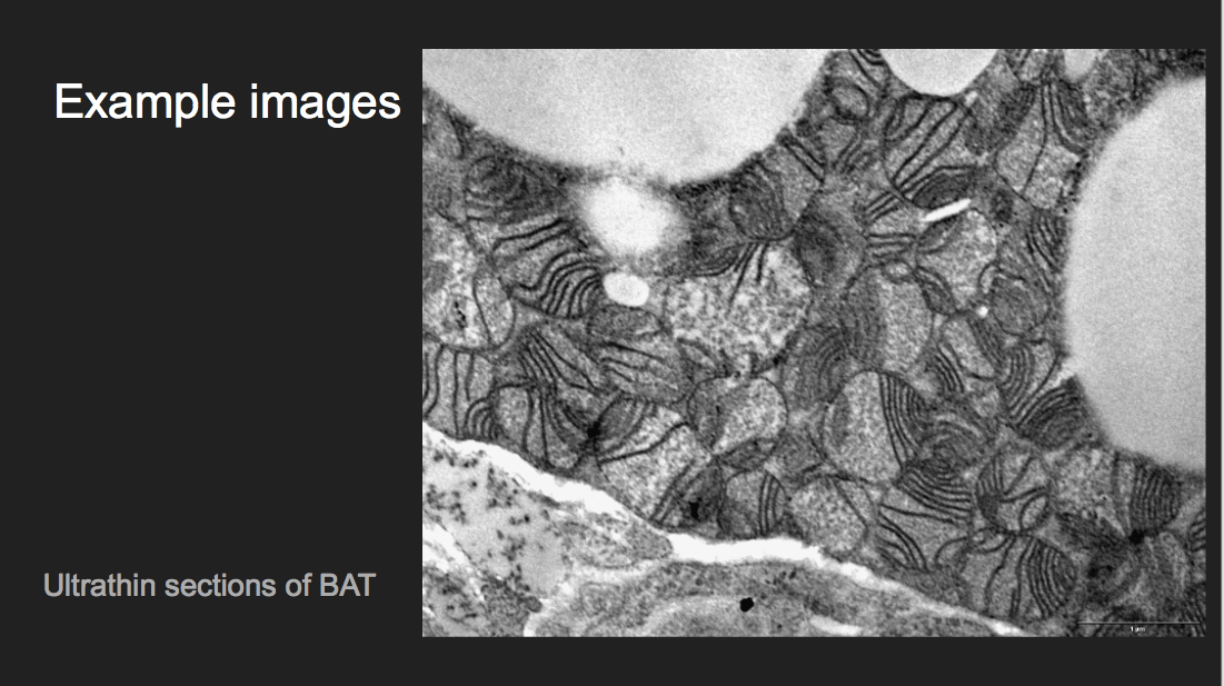
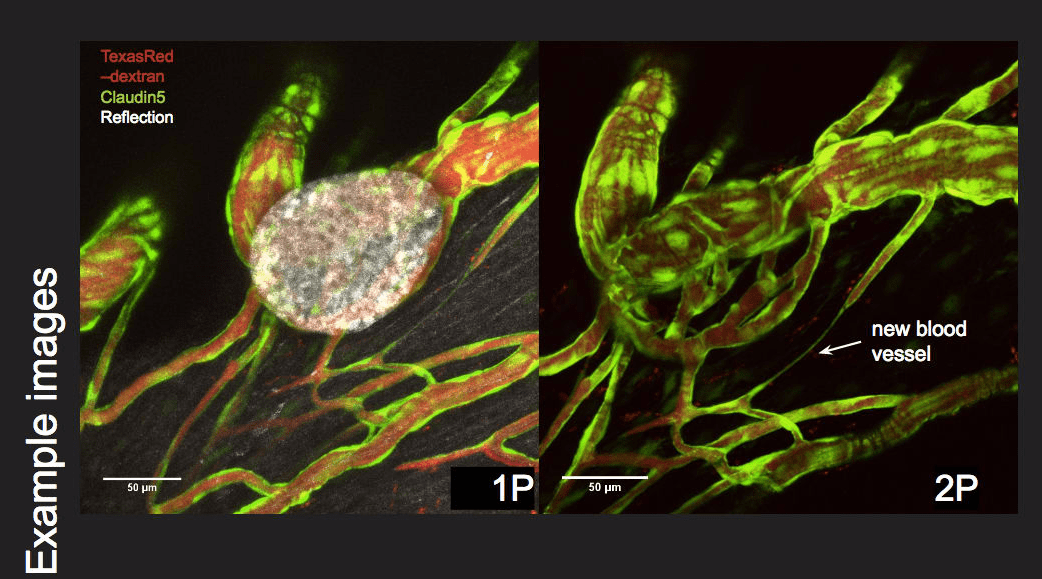
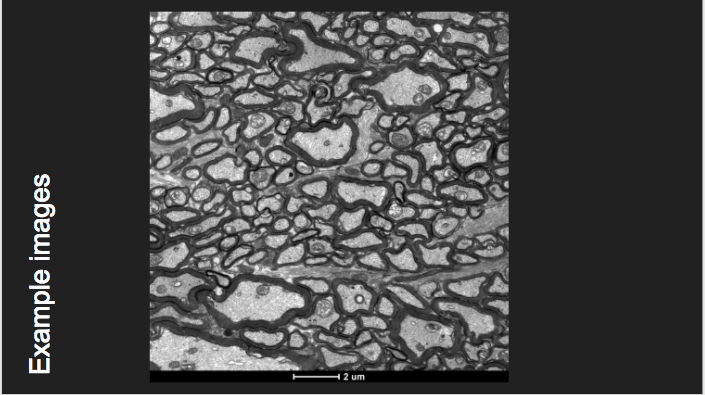
Cases
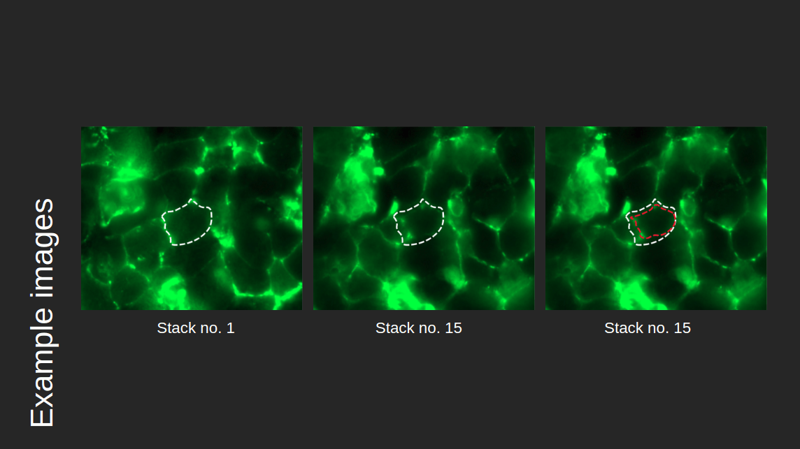
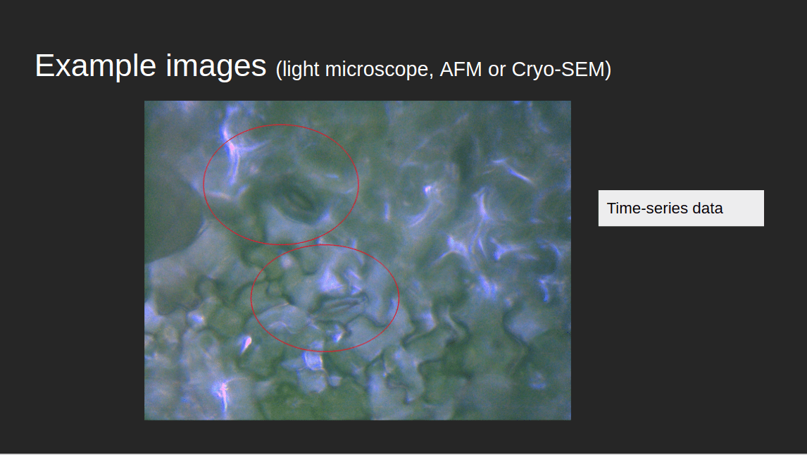
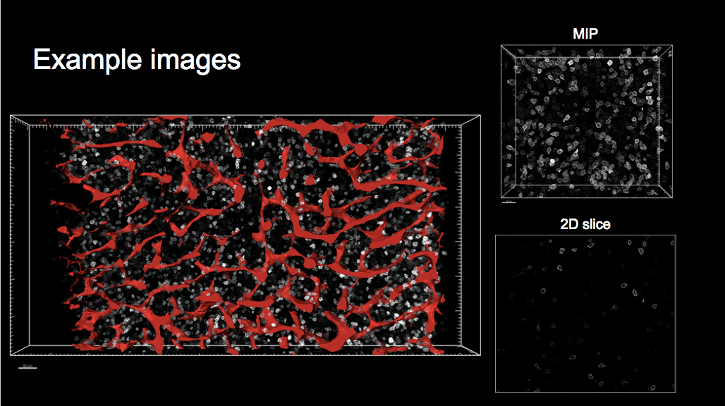
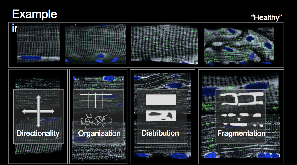
Watch presentations and share your insights

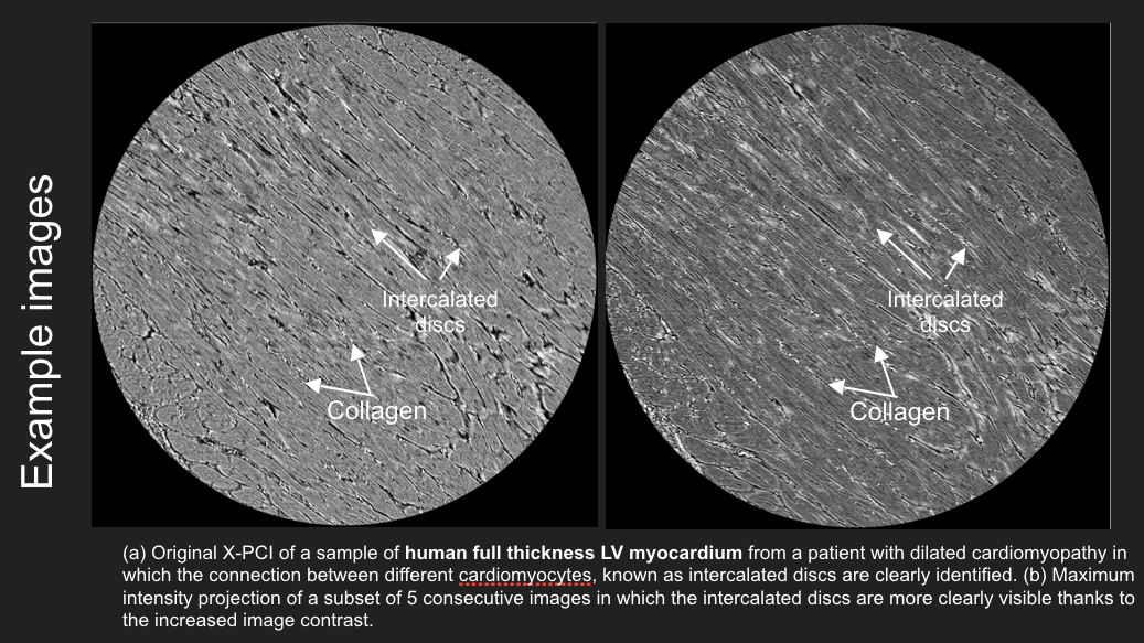
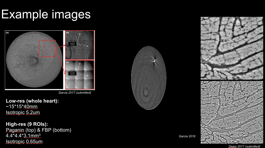
Quantification of cardiac micro-structures on intact whole heart using synchrotron X-ray imaging
Team: Chong Zhang, Patricia Garcia-Canadilla, Hector Dejea, Anne Bonnin, Vedrana Balicevic, Marco Stampanoni, Bart Bijnens, Andrew C. Cook
Resources
Quick links


Events
Events where Call4Help session are organized

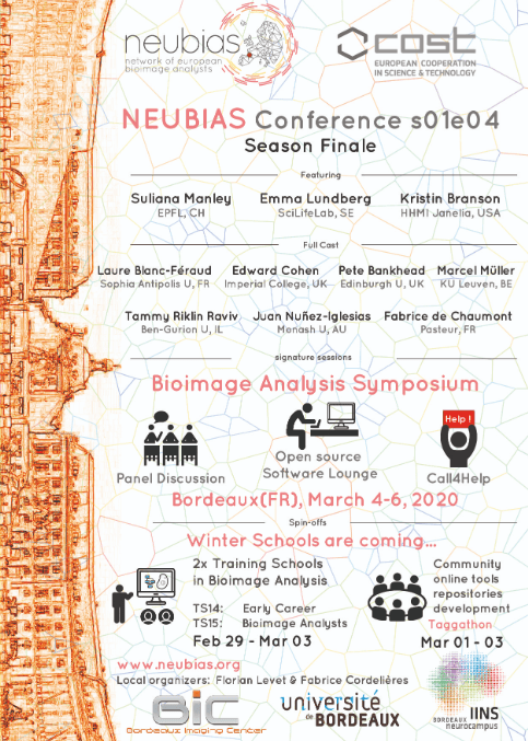
4th NEUBIAS Conference
4th - 6th Feb. 2020, Bordeaux
The 2nd Annual Conference of NEUBIAS, gathering the whole BioImage Analysis Community into a multi-faceted event, offers two Training schools, a Working Meeting (Taggathon) and a Large Symposium dedicated to Scientific Developments and Open Tools in BioImage Analysis. [more about the conference]

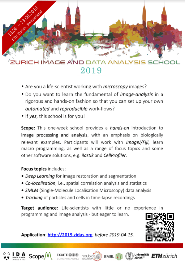
ZIDAS 2019
18-23 Jun. 2019, Zurich
This one-week school provides a hands-on introduction to image processing and analysis, with an emphasis on biologically relevant examples. You will learn the fundamentals of image analysis, including basic macro programming in ImageJ/Fiji as well as other software solutions. In the first part of the week we will also cover the process of image-formationas it pertains to image analysis: Resolution, correct exposure, point-spread functions, detector noise, Shannon's sampling theorem, and aliasing. All with a clear focus on application in the lab. [more about the conference]
Past events

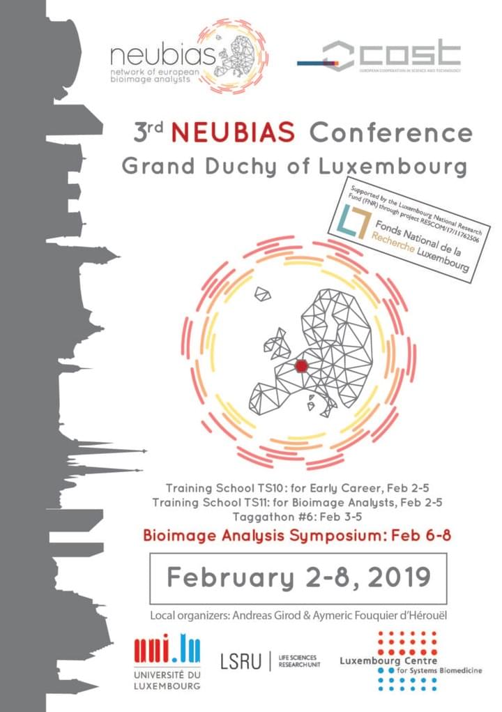
3nd NEUBIAS Conference
31st Jan. - 2nd Feb. 2019, Luxemburg
The 3rd Annual Conference of NEUBIAS, gathering the whole BioImage Analysis Community into a multi-faceted event, offers two Training schools, a Working Meeting (Taggathon) and a Large Symposium dedicated to Scientific Developments and Open Tools in BioImage Analysis. [more about the conference]

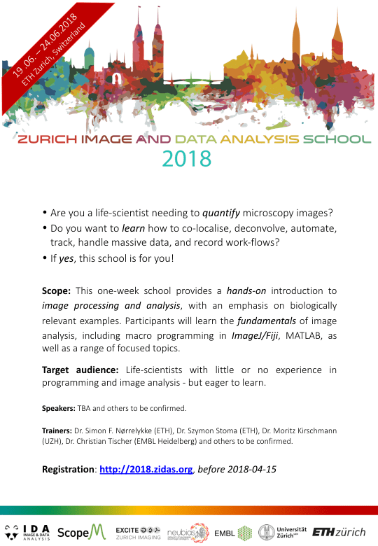
ZIDAS 2018
16th - 20th June 2018, Switzerland
This one-week school provides a hands-on introduction to image processing and analysis, with an emphasis on biologically relevant examples. You will learn the fundamentals of image analysis, including basic macro programming in ImageJ/Fiji as well as other software solutions. In the first part of the week we will also cover the process of image-formationas it pertains to image analysis: Resolution, correct exposure, point-spread functions, detector noise, Shannon's sampling theorem, and aliasing. All with a clear focus on application in the lab. [more about the conference]

2nd NEUBIAS Conference
31st Jan. - 2nd Feb. 2018, Hungary
The 2nd Annual Conference of NEUBIAS, gathering the whole BioImage Analysis Community into a multi-faceted event, offers two Training schools, a Working Meeting (Taggathon) and a Large Symposium dedicated to Scientific Developments and Open Tools in BioImage Analysis. [more about the conference]

ZIDAS 2017
16th - 20th October 2017, Switzerland
This one-week school provides a hands-on introduction to image processing and analysis, with an emphasis on biologically relevant examples. You will learn the fundamentals of image analysis, including basic macro programming in ImageJ/Fiji as well as other software solutions. In the first part of the week we will also cover the process of image-formationas it pertains to image analysis: Resolution, correct exposure, point-spread functions, detector noise, Shannon's sampling theorem, and aliasing. All with a clear focus on application in the lab. [more about the conference]

BIAS
14-20 May 2017, Germany
Organised by EMBL, the topics of this conference reflect the scientific research carried out by EMBL scientists or bring together long established communities sometimes outside the realm of EMBL research. This platform allows for the exchange of our exciting findings with the international scientific community.

NEUBIAS2020
12-17 Feb. 2017, Portugal
NEUBIAS2020 conferences bring together Life-Scientists, Bioimage Analysts, Image Analysis- and Software- developers and other Core Facilities Specialists (e.g. Microscopists) to bridge the gap between Bioimage Analysis developments and know-how, and their use in Life Science. It aims to be a forum to exchange the newest findings, applications and cutting-edge developments in Bioimage Analysis, machine learning, data mining and storage.
Support
Organization supporting us